
Parasternal longaxis view demonstrating pericardial effusion and
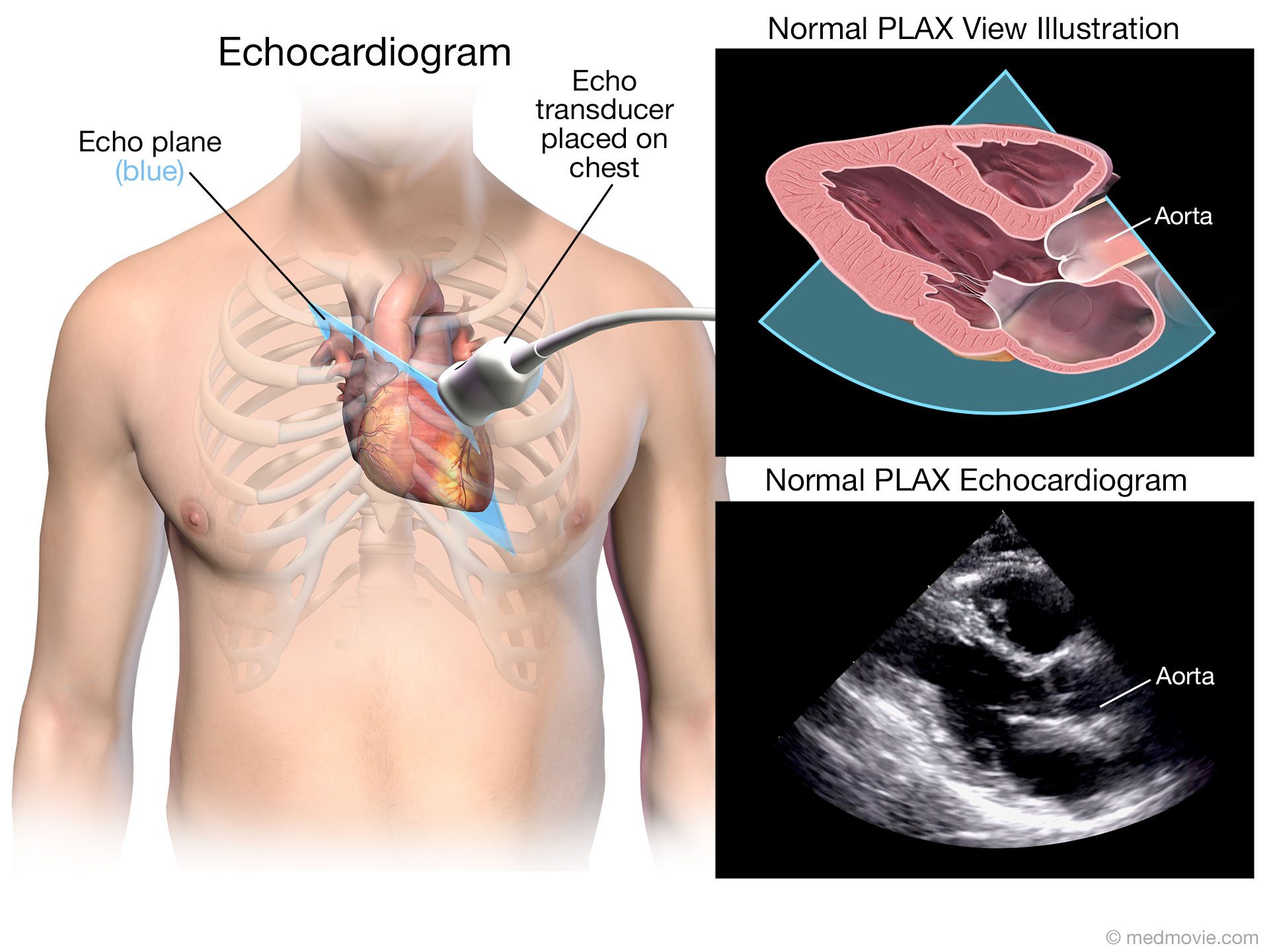
Parasternal long axis (PLAX) view is one of the easiest views to obtain and answers most of the questions encountered in day-to-day nephrology practice. The sonographic anatomy, image acquisition and key pathologies seen in this view are discussed below. How is the exam performed? The transducer or the probe:
Basic echocardiographic views All About Cardiovascular System and
The basic concept of echocardiography was first demonstrated by Lazzaro Spallanzani in the 18 century when he described the reflected echoes of inaudible sound. [1] Hertz and Edler reported the first use of ultrasound in the cardiac evaluation and continuous monitoring of heart movements in 1954. [2]

Dr.Nabil Paktin's Journal of Cardiovascular Medicine Blog ژورنال ( قلب
The segments are studied in six views: the parasternal long axis, the parasternal short axis at the levels of the mitral valve, papillary muscles, and apex, apical four chambers, apical two chambers. The scoring system is based on if the wall motion is normal, hypokinetic, akinetic, or dyskinetic. Based on the wall motion, a score of 1 to 4 is.

Transthoracic echocardiogram in a parasternal long axis window The BMJ
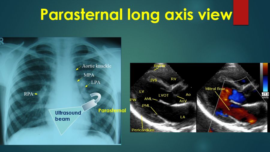
Parasternal Long-Axis View . From the parasternal position, the probe should be adjusted so that the transducer orientation marker is pointing toward the patient's right shoulder ( Figure 13.4 ). The ultrasound beam should be positioned parallel to a line running from the patient's right shoulder to their left hip. Images obtained represent.

The parasternal longaxis view (A) and shortaxis view (B) of an
In this paper, we will outline the standard and conventional parasternal long and short axis views. Figure 1 Surface projections of the heart (red), pleurae (light blue) and lungs (violet) The study commences with the parasternal long axis view (PLAX - figure 2 ). Figure 2 Schematic view of parasternal long axis ultrasound beam Figure 3

Cardiac Transthoracic Echocardiography (TTE) Summary And Labeled
The most common cross-sectional views are the parasternal long axis, the parasternal short axis, and the apical view (see Figure 1 ). The gastric or subcostal and suprasternal views are also commonly used. Figure 1 The most common two-dimensional imaging echo views.
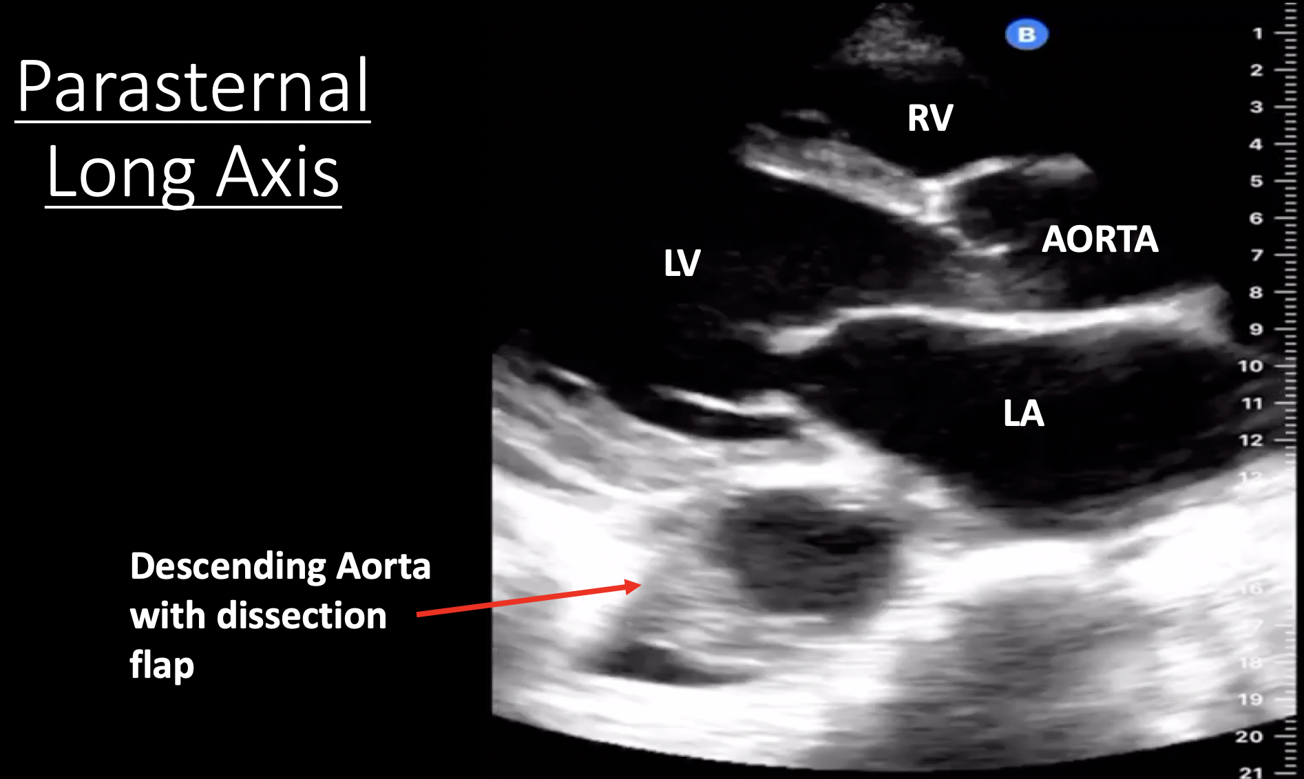
Aortic Dissection Annotated Still Image 1, Parasternal Long
Video 3. Parasternal long axis-view; Abdominal and Lower Thoracic Views. When a patient is in the supine position the most dependent area in the upper peritoneum between the liver and right kidney, also known as Morison's pouch, and the most dependent area in the lower peritoneum is posterior to the bladder in the male and the pouch of Douglas (posterior to the uterus) in the female.

Parasternal long axis view of a representative echocardiographic image
The parasternal long-axis view (PLAX) is located on the left side of the sternum. It provides imaging planes of the long axis of the heart. Figure 2 illustrates the position of the transducer, the orientation of the index marker and the scanning plane through the heart.
FileTransthoracic Echo Parasternal Long Axis LV Schematic.png Wikipedia
The parasternal long axis (PLA) is the first image in a transthoracic echocardiogram (TTE). It is an important window because it allows assessment of the left ventricular ejection fraction (LVEF) and measurement of the LV outflow tract diameter (LVOTD). The PLA can be hard to obtain in ICU patients.
Parasternal Long Axis
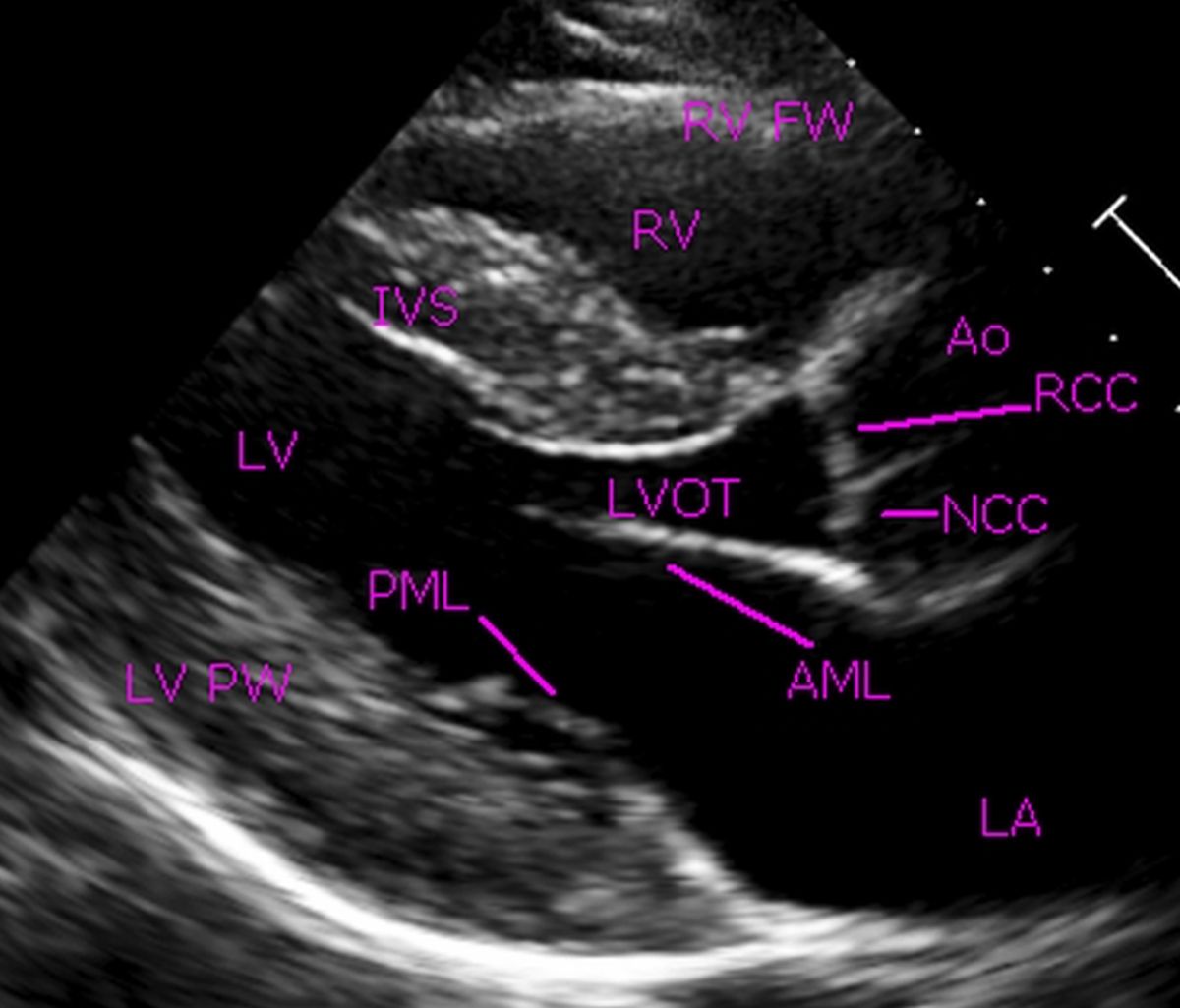
Parasternal long-axis view with the origin of the right coronary artery The PLAX view also permits measurement of the size of the left atrium (especially in its anterior/posterior extension) and is also very important for the interpretation of valvular function.

1A Parasternal long axis view of prolapsus of anterior and posterior
Parasternal Long-Axis View. A pericardial effusion is seen as an anechoic (black) region between the hyperechoic (bright) pericardium and the walls of the heart. The image demonstrates a small pericardial effusion, while the illustration demonstrates the location of a larger (circumferential) effusion.
Parasternal long axis view in normal echocardiogram
Normal parasternal long axis view; Parasternal short axis view: This view is a cross sectional view of the left and right sides of the heart. These can be "sliced" at various levels between the base and the apex. By fanning the probe towards the right shoulder, one can visualize the aortic valve in cross section.

Pin on Cardiac surgery
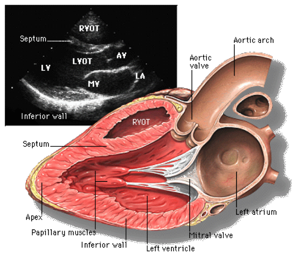
Standard Parasternal Long Axis (PLAX) Landmarks Right ventricle or right ventricular outflow tract Left ventricle, aortic valve and proximal aorta Mitral valve and left atrium

2.3.1 Parasternal window Longaxis views (PLAX) 123 Sonography
sional (2D) imaging (Figure 5). Alternatively, the left parasternal view is also used for measuring RV wall thickness. Thickness > 5 mm indicates RV hypertrophy (RVH) and may suggest RV pressure overload in the absence of other pathologies. IVC DIMENSION. The subcostal view permits imaging and measure-
Parasternal long axis view of the transthoracic echocardiography which
The Parasternal Long Axis View is often abbreviated as PSLA or PLAX. It is usually the first cardiac ultrasound view obtained and will give you an immediate assessment of the general condition of the heart including ejection fraction and overall left and right ventricular sizes.
Making sense of an echocardiogram report for GPs! — Cardiology Institute
Transthoracic echocardiography (TTE) is the primary initial imaging modality in cardiac imaging. Advantages include portability, safety, availability, and ability to assess the morphology and physiology of the heart in a noninvasive manner.